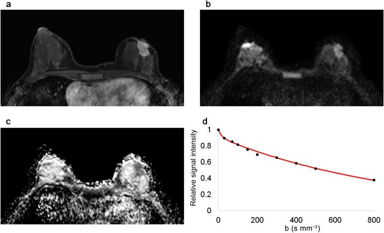
Figure 2.
Magnetic resonance images of a 46-year-old female with luminal A type invasive ductal carcinoma in the left breast. The malignant mass is shown in (a) a contrast-enhanced T1 weighted image, (b) a diffusion-weighted image (b = 800 s mm−2) and (c) an apparent diffusion coefficient map. (d) The graph shows the biexponential signal decay depending on the b-value.

