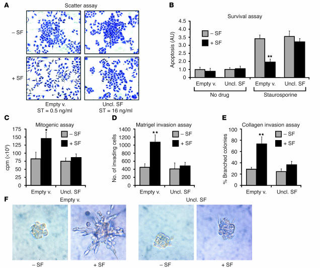
Figure 4.
Uncleavable SF inhibits the pleiotropic activities of SF on living cells. (A) End-point scatter assay. A549 cells transduced with the indicated lentivirus vector were stimulated with progressive 1:2 dilutions of active SF starting from 64 ng/ml. The minimal concentration at which scattering was observed is indicated (scatter threshold [ST]). Representative images show crystal violet–stained cells at 8 ng/ml (the concentration of active SF). (B) Survival assay. Lentivirus vector–transduced MDA-MB-435 cells were pre-incubated with either recombinant SF (+SF) or no factor (–SF) and then were cultured in the absence (No drug) or presence (Staurosporine) of staurosporine. (C) Mitogenic assay. Lentivirus vector–transduced A549 cells were stimulated with either recombinant SF or no factor, and DNA synthesis was assessed by [3H]thymidine incorporation. (D) Matrigel invasion assay. MDA-MB-435-β4 cells were analyzed for their ability to invade a Matrigel layer in the presence or absence of recombinant SF. (E) Collagen invasion assay. Lentivirus vector–transduced MDA-MB-435 cells were examined for their ability to form branched, multicellular tubules in response to SF stimulation. (F) Representative images from the experiment described in E. Statistical significance (*P < 0.05; **P < 0.01) refers to the difference between –SF and +SF.

