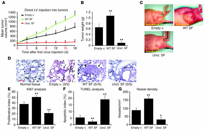
Figure 7.
Uncleavable SF arrests the progression of established tumors. (A) Tumor burden analysis. Tumors of the same size (approximately 100 mm3 at day 0) were repeatedly injected with the indicated lentivirus vectors, and tumor volume was measured every 3 days. The numbers in red correspond to the days of lentivirus injection. LV, lentivirus vector. (B) Tumor weight analysis for the experiment described in A. (C) Representative images of experimental tumors. The yellow ruler indicates length in centimeters. (D) Histological analysis of pulmonary metastases on lung sections stained with hematoxylin and eosin. A representative microscopic field for each group is shown. Arrows indicate the position of the metastatic lesion. Black staining is due to India ink. Metastasis incidence is shown in parenthesis. Red scale bar: 100 μm. (E) Tumor proliferation index analysis (% Ki67-positive cells). (F) Tumor apoptotic index analysis (% TUNEL-positive cells). (G) Tumor vessel analysis using the CD31 endothelial marker. Vessel density, number of vessels/mm2. Statistical significance was calculated as for Figures 5 and 6.

