
Figure 2.
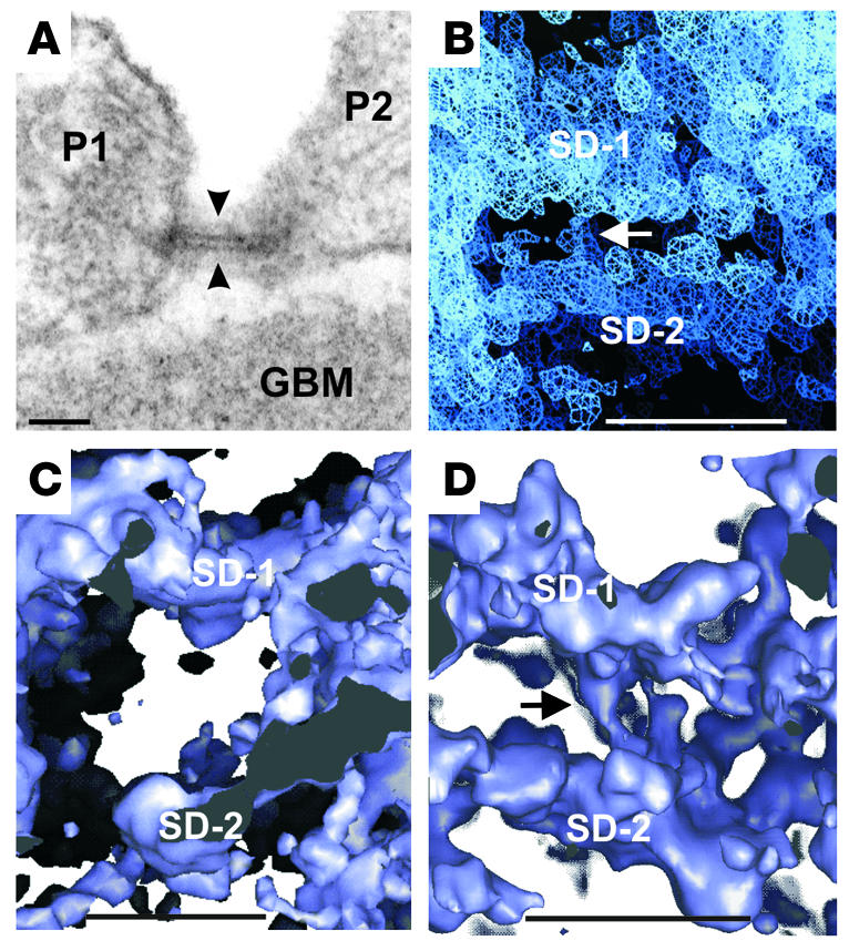
Human, rat, and mouse slit diaphragm with double layers. Scale bars: 50 nm (A), 20 nm (B–D). (A) Filtration slit in EM cross section between foot processes of human podocytes (P1, P2) showing double-layered slit diaphragm (arrowheads). Tannic acid–glutaraldehyde and osmium fixation; resin section. (B) Tomogram of same slit as in A. Strands seem to connect the two slit diaphragm layers (arrow). Sigma level: 0.5. (C) Same rat filtration slit as in Figure 1F, tilted 90– around the x axis; showing 2 layers in slit diaphragm. Sigma level: 1.0. (D) Two layers in cross-cut mouse slit diaphragm with connecting strands (arrow); glutaraldehyde and osmium fixation. Sigma level: 1.0.