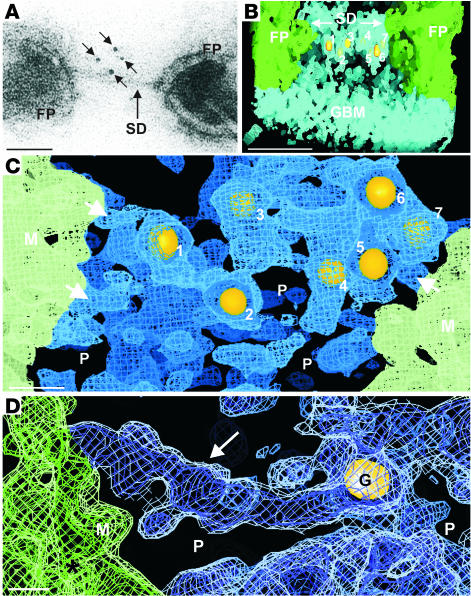
Figure 3.
Localization of nephrin in human slit diaphragm: immuno-cryolabeling of extracellular terminal Ig-domains of nephrin. SD pores (P) are indicated. Scale bars: 40 nm (A and B), 10 nm (C), 5 nm (D). (A) Nephrin label (5-nm gold, arrows) in EM along obliquely cut slit diaphragm. (B) Tomogram of filtration slit bordered by GBM, foot processes, and slit diaphragm. Gold labeling for nephrin appears under the diaphragm at different levels of the digital volume. Sigma levels: 0.05 (tissue), 13 (gold particles). (C) Higher magnification of B (same sigma levels), but visualized from below, through 30-nm-thick digital section encompassing the slit diaphragm. Around 4-nm-wide strands (arrows) extend from the podocyte surface into the diaphragm. (D) Close-up of slit diaphragm cross strand (arrow). Gold label (G) appears near the distal end of cross strand. Note associated globules (sectioned short strands) at the proximal part of cross strand. Only small volume differences in wire frames exist between sigma levels 1.0 (blue and green) and 0.3 (white).

