Figure 4.
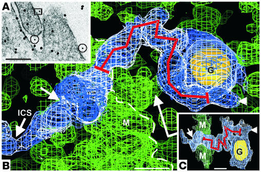
Extracellular nephrin-label on transfected HEK293 cells. Scale bars: 100 nm (A), 5 nm (B and C). (A) Small, 5-nm-immunogold particles (in the rectangle and 2 circles) mark nephrin on the cell surface. (The large 10-nm-gold particles are used as coordinates for 3D-reconstruction purposes [39, 67].) Pre-embedding immunolabeling; resin section. (B) Tomogram from reconstructed volume of rectangle in A. The strand with a gold label on its distal end seemingly traverses the cell membrane. Marked extracellular length, measured in 3D, is about 35 nm when the putative anti-nephrin IgG complex (5-nm-gold–anti-rabbit IgG + rabbit anti-nephrin IgG) at the end of the strand (arrowhead) is omitted. Inside the cell, the strand is continuous near the membrane (short arrow) with intracellular strand (ICS). Sigma levels: 0.5 (green and blue) and 0 (white, strand-immunogold complex); 13 (gold particle). (C) A 90–-tilted side view from the direction shown in B (long, bent arrow). Sigma levels: 0.3 (green and blue) and 0 (white). Note minimal volume change in strand between sigma levels in B (from 0.5 to 0) and C (from 0.3 to 0).

