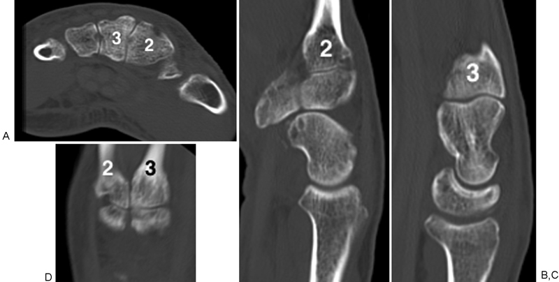
Fig. 5.

A 71-year-old man with right-sided pisiform and triquetral fractures following ground level fall. Axial (A), sagittal (B and C), and coronal (D) unenhanced CT imaging at the level of the base of the second and third CMC joints. There is no measurable dorsal protuberance at the base of the second or third metacarpal, no separate dorsal ossicle at the second or third CMC joint, and no dorsal protuberance arising from the trapezoid or capitate. (2 = 2nd metacarpal; 3 = 3rd metacarpal). CMC, carpometacarpal; CT, computed tomography.
