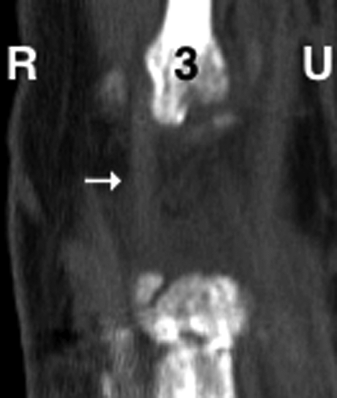
Fig. 6.

A 65-year-old woman with left-sided distal radius fracture following open reduction and internal fixation. Coronal unenhanced CT image at the dorsal base of the third metacarpal demonstrates attachment of the ECRB (arrow) to the radial and dorsal margins about the protuberance. (R = radial side; U = ulnar side; 3 = 3rd metacarpal). CT, computed tomography; ECRB, extensor carpi radialis brevis.
