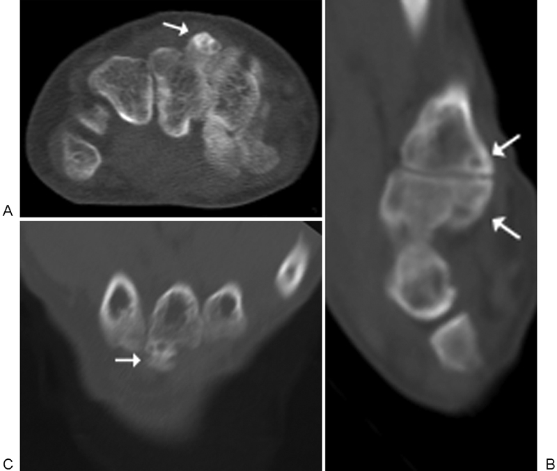
Fig. 7.

A 53-year-old man with left-sided distal radius fracture. Axial (A), sagittal (B), and coronal (C) unenhanced computed tomography images at the third carpometacarpal joint demonstrate a dorsal protuberance from the base of the third metacarpal with an adjacent protuberance from the capitate, forming a pseudoarticulation. Changes suggestive of secondary arthritis are present (arrows).
