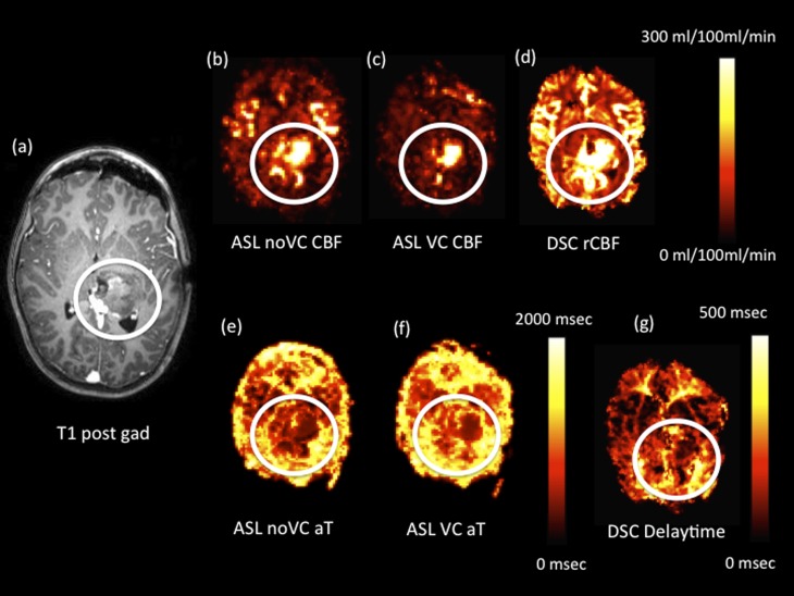
Figure 4.
Intraoperative axial images from a single patient following a tumour-debulking surgery of a high-grade anaplastic astrocytoma (the region of residual tumour tissue is highlighted in the white circles). (a) Axial T1 weighted post-gadolinium (post gad) image. Cerebral blood flow (CBF) images for arterial spin labelling (ASL) with no vascular crushing (i.e. ASL noVC) (b), ASL with vascular crushing (c) and relative cerebral blood flow (rCBF) dynamic susceptibility contrast (DSC) (d) are shown with different scales. Arterial arrival time (aT) ASL with vascular crushing/delay time images for ASL (e), ASL with vascular crushing (f) and delay time from DSC (g) are shown with different scales. VC, vascular crushers.

