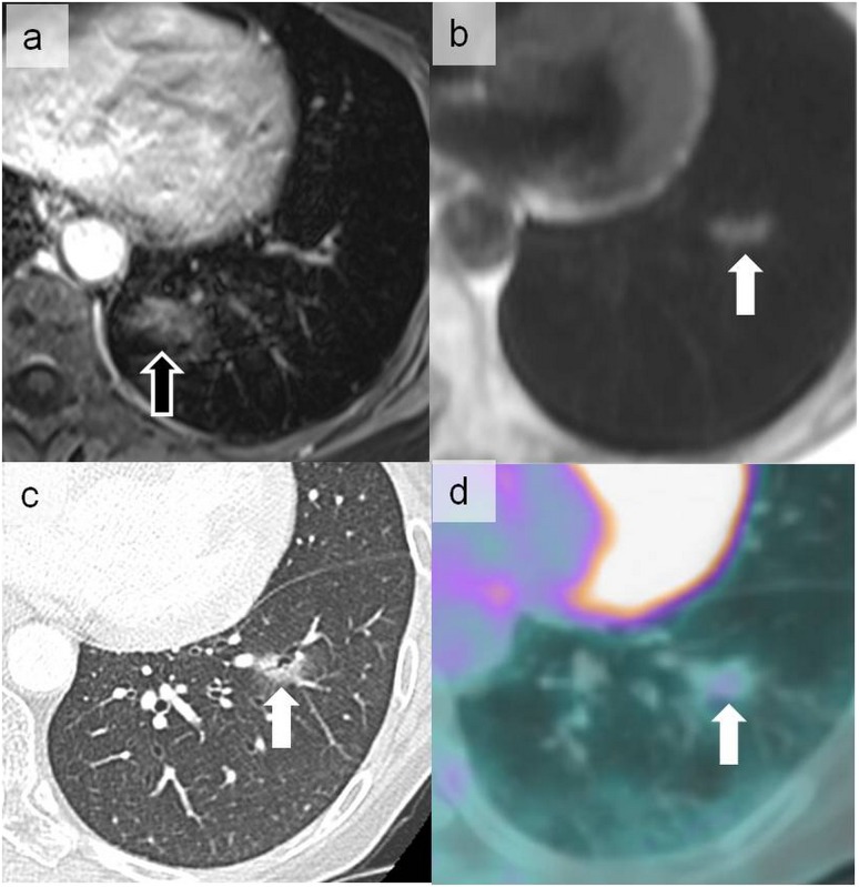
Figure 15.
Cardiac ghost artefacts on the volumetric interpolated breath-hold examination sequence more prominent in the phase-encoding direction projecting posteriorly on the lung parenchyma (open arrow) (a). This should not be confused with the abnormal signal more laterally located in the left lower lobe on a half-Fourier acquisition single-shot turbo spin-echo sequence (solid arrow) (b). The latter signal corresponded to a ground-glass nodule on CT (c) metabolically hyperactive on positron emission tomography (d) that was related to pulmonary localization of a primary gastric lymphoma (arrows).

