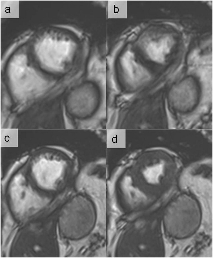
Figure 17.
End-diastolic (a) and end-systolic (b) cine images in a patient with atrial fibrillation (irregular R–R interval) using retrospective electrocardiogram (ECG) gating. With this method, the images are acquired continuously throughout the cardiac cycle over several (irregular) heart beats. Using prospective ECG triggering, the acquisition window is limited to the systolic phase (in this case, 350 ms after R-wave), partially avoiding the artefacts due to irregular R–R interval. This approach results in a better image quality at end-diastole (c) and end-systole (d) and throughout the imaged cardiac cycle.

