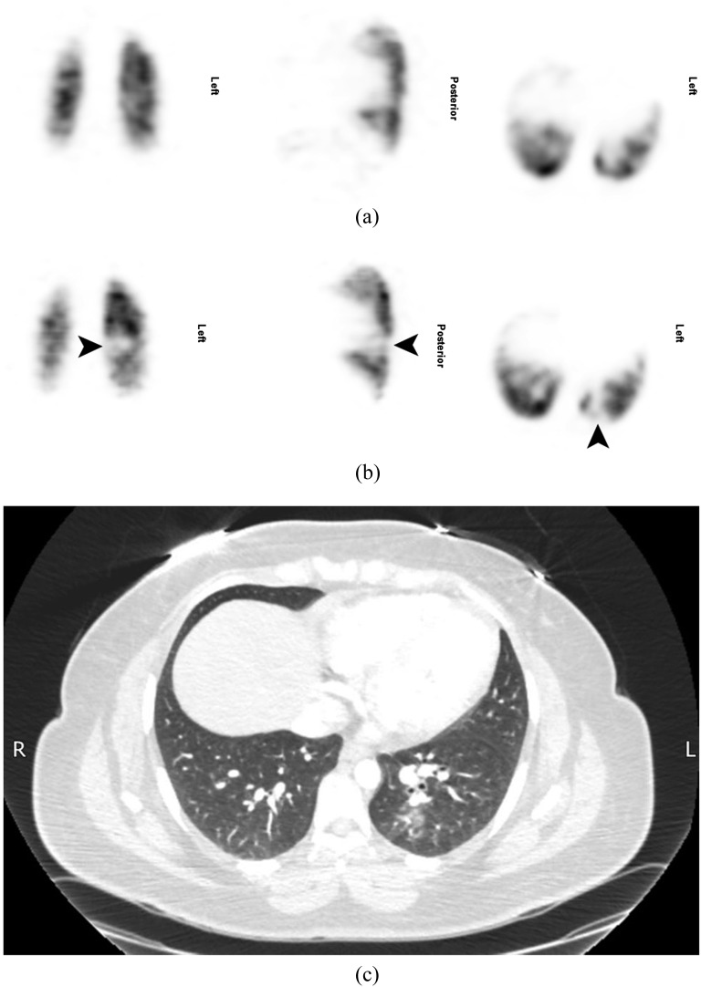
Figure 3.
A 30-year-old female who was 15 weeks pregnant presented with a sudden onset of pleuritic chest and back pain, a cough and small amounts of haemoptysis. She had a normal chest X-ray at presentation and a normal Doppler scan of the lower legs 3 days later (not shown). A lung perfusion single-photon emission CT (SPECT) was performed the same day (b), and the patient was recalled for a ventilation SPECT the following day (a). This showed a singular subsegmental mismatched perfusion defect at the left base (arrows) and the scan was reported as indeterminate. The same day, CT pulmonary angiography (c) achieved good contrast opacification (378 HU in the main pulmonary artery), but no pulmonary embolism was seen, although there was ill-defined nodularity and patchy ground-glass opacification in the superior and posterior segments of the left lower lobe in keeping with infection.

