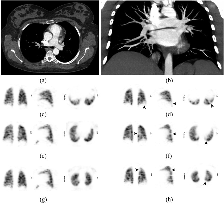
Figure 4.
A 26-year-old female who was 38 weeks pregnant presented with a 1-day history of right upper pleuritic back pain and some shortness of breath. She had a normal chest X-ray at presentation and a normal Doppler scan of the lower legs on the following day (not shown). The patient received anticoagulation with clexane; a lung perfusion single-photon emission CT (SPECT) was performed another day later (c, e, g, ventilation images; d, f, h, perfusion images), and she was recalled for a ventilation SPECT the following day. This showed two or possibly three subsegmental mismatched perfusion defects in the left lung (arrows) (a, transverse view; b, maximum intensity projection) in keeping with pulmonary embolism (PE). CT pulmonary angiography another 2 days later achieved good contrast opacification (375 HU in the main pulmonary artery), but no PE was seen.

