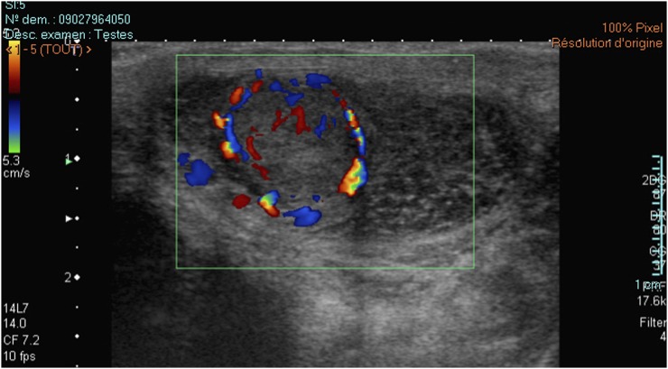
Figure 5.
A typical Leydig cell tumour (LCT) as seen in a patient presenting with Klinefelter syndrome (KS). Note the small testis volume and coarse hypoechoic echotexture typically seen in patients with KS. The LCT appears as a large hyperechoic heterogeneous mass in B-mode sonography and presents marked hypervascularization with a central corbelling pattern.

