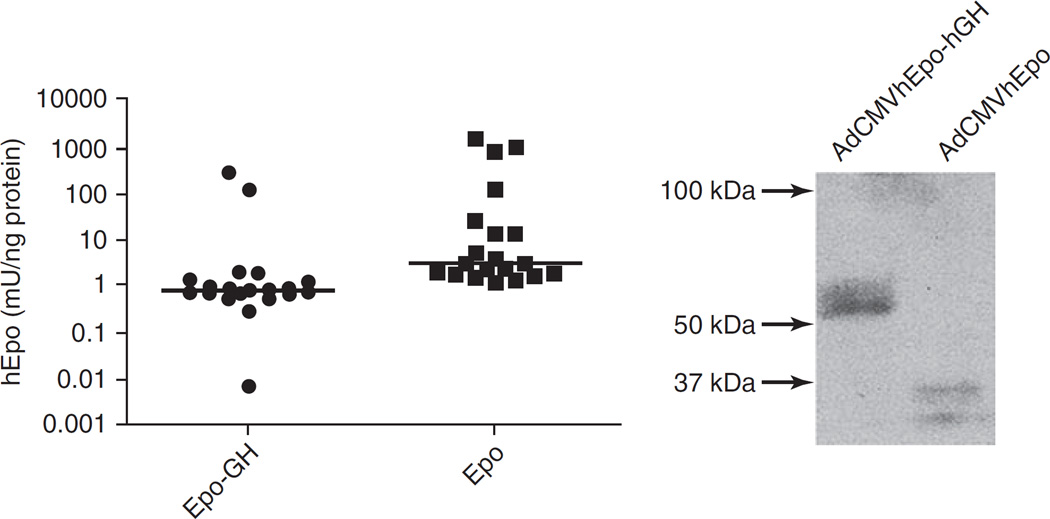
FIG. 3.
Immunoreactive hEpo in aqueous extracts of mouse submandibular glands. Left: Forty-eight hours after administration of AdCMVhEpo-GH or AdCMVhEpo at 1010 viral particles/gland, glands were harvested and frozen at −80°C. Thereafter, glands were homogenized and levels of immunoreactive hEpo were determined in gland extracts by ELISA. Glands transduced with AdCMVhEpo had a median protein level of 2.99 mU/ng protein (range, 1.18–1292.4 mU/ng protein), whereas glands transduced with AdCMVhEpo-hGH showed a median protein level of 0.79 mU/ng protein (range, 001–255.6 mU/ng protein). The horizontal bar represent the median value (n = 20, p < 0.001). Right: Extracts from glands were electrophoresed as described in Materials and Methods, blotted, and then reacted with anti-hEpo antibody. Mouse glands transduced with AdCMVhEpo-hGH or AdCMVhEpo expressed immunoreactive hEpo–hGH and hEpo protein bands with apparent molecular sizes of ~56–58 and ~32–34 kDa, respectively.

