FIGURE 4.
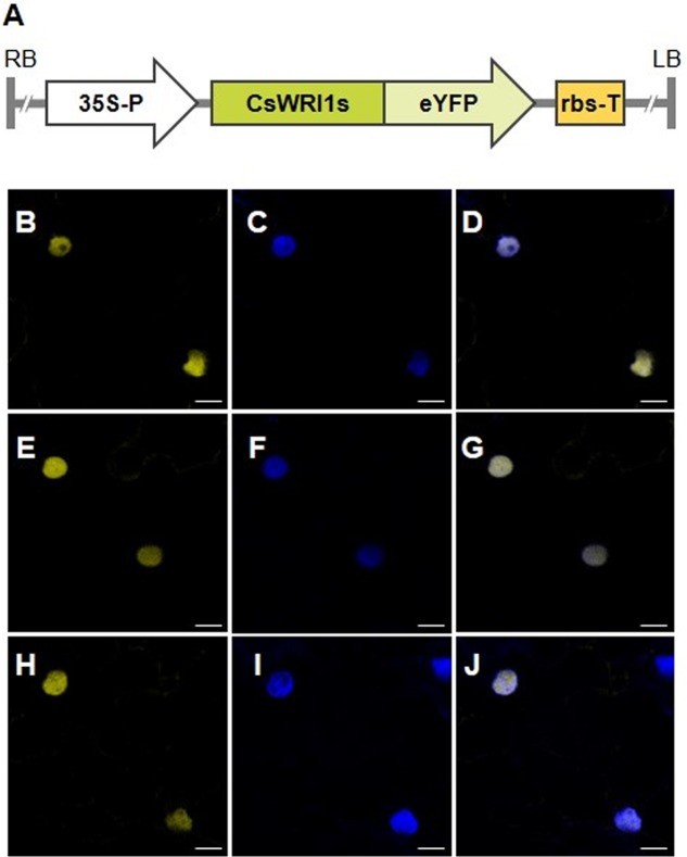
Subcellular localization of CsWRI1s:eYFP proteins in N. benthamiana epidermis. (A) Schematic diagram of CsWRI1A:eYFP. CsWRI1B:eYFP, and CsWRI1C:eYFP constructs. 35S-P, cauliflower mosaic virus 35S promoter; LB, left border; RB, right border; rbs-T, the terminator of ribulose-1,5-bisphosphate carboxylase and oxygenase small subunit from pea (Pisum sativum). (B–J) Agrobacterium harboring the CsWRI1A:eYFP (upper row), CsWRI1B:eYFP (middle row) or CsWRI1C:eYFP (bottom row) construct was infiltrated into N. benthamiana leaves and then the fluorescent signals were visualized under laser confocal scanning microscopy. YFP signals (B,E,H) from the CsWRI1s:eYFP constructs. The nucleus (C,F,I) was visualized by staining with DAPI under the UV filter. Merged image between signals of YFP and the nucleus (D,G,J). EV, empty vector (pBA002). Bars = 10 μm.

