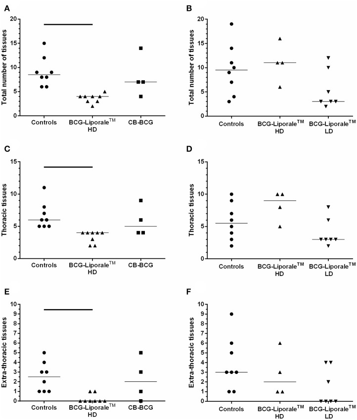
Figure 3.
Vaccination of badgers with BCG and challenged 13 weeks later with endobronchial M. bovis. Number of organs/tissues from which M. bovis was isolated or AFB found 12 weeks post-challenge for VES3 (left panel) and VES4 (right panel). The total number of affected tissues is shown (A,B), together with their distribution: thoracic (C,D) or extra-thoracic (E,F). Individual animal results are shown together with the group median. Badgers were vaccinated with either CB-BCG (■), HD BCG-Liporale™ (▲) or LD BCG-Liporale™ vaccine (▼). Liporale™ alone was used as a negative control for vaccination (•). A significant difference according to Dunn's test for multiple pair wise comparisons is shown by the bar.

