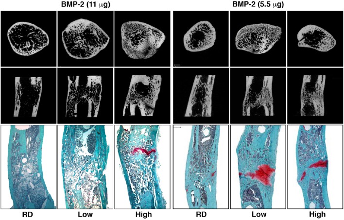
Figure 2.
Micro-computed tomography (μCT) and histology images illustrating healing of segmental defects with 11 and 5.5 μg doses of BMP-2, high and low stiffness external fixator groups as well as a Reverse Dynamization group (RD) after 8 weeks of treatment. μCT images of cross-sectional distal part of the defect (top row) and coronal plane of the defect (middle row). Histological sections were stained with safranin orange-fast green (bottom row, Scale bar = 1 mm). Low = low stiffness fixator (114 N/mm); High = high stiffness fixator (254 N/mm); RD = low to high stiffness fixation at 2 weeks (from Glatt et al., 2012b, 2016a).

