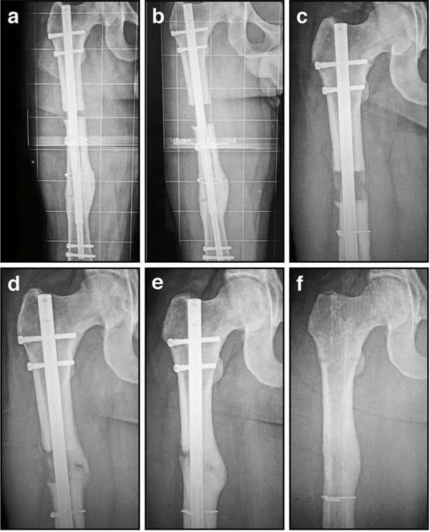
Figure 6.

Radiographic images illustrating the process of gradual femoral lengthening using a telescopic nail: (A) AP radiograph of a femoral lengthening nail obtained early post-operative, following only limited distraction; (B) AP radiograph of a telescopic lengthening nail after completing distraction of 2.7 cm; (C) Radiographic image illustrating lengthening gap at 6 weeks, demonstrating early regenerate bone formation; (D) Radiographic image at 12 weeks, as the regenerate gradually matures and consolidates; (E) Radiographic image illustrating lengthening gap at 24 weeks, as the regenerate bone matures further and hypertrophies; (F) Radiographic image illustrating fully consolidated gap at 52 weeks, following removal of the telescopic nail.
