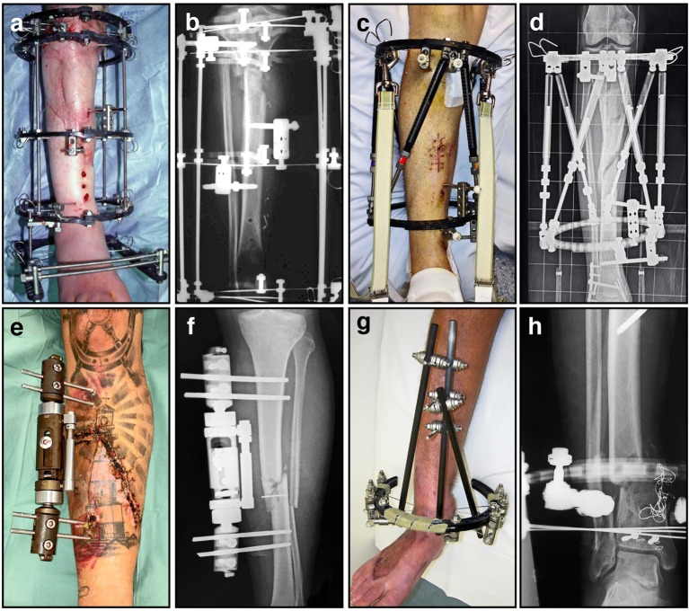
Figure 7.
A variety of external fixators are used for fracture fixation, most often for tibia fractures as demonstrated here. (A) Clinical image of an Ilizarov external fixator applied to a fractured leg, with multiple rings and tensioned wires; (B) Corresponding radiograph illustrating proximal tibial fracture fixation with this circular frame; (C) Clinical image of a hexapod-based external fixator applied to a fractured limb, using a pair of rings; (D) Corresponding radiograph illustrating stabilization of a comminuted segmental tibia fracture; (E) Clinical image of a unilateral fixator in position; (F) Corresponding radiograph demonstrating mid-tibial open fracture fixation using this device; (G) Clinical image of a hybrid (cantilever) external fixator, incorporating a single juxta-articular ring and unilateral diaphyseal elements; (H) Corresponding radiograph demonstrating a distal tibial fracture stabilized using this device.

