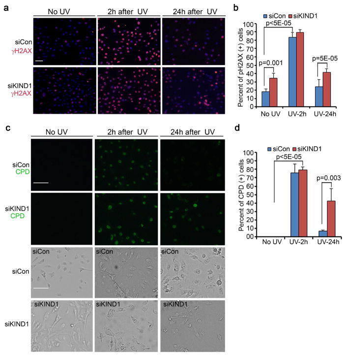
Figure 3. KIND1-loss sensitizes keratinocytes to UVB-induced DNA-damage.
(a) Immunofluorescent staining of γH2AX. Human keratinocytes transfected with siCon or siKIND1 were treated with UVB for 5 seconds (6.8mJ/cm2), and then immunostained for γH2AX followed by detection with an Alexa-555-conjugated secondary antibody. γH2AX [orange], nuclei [blue, Hoechst]. (b) Quantification of γH2AX-positive cells. Graph represents average percentages of γH2AX-positive cells + SD. (c) Immunofluorescent staining with a FITC-conjugated antibody against CPD [green]. Bright field images were shown below each corresponding CPD image. (d) Quantification of CPD-positive cells. Graph represents average percentages of CPD-positive cells + SD. P-values of <0.05 were obtained with student T-test. Scale bars: 50 μm.

