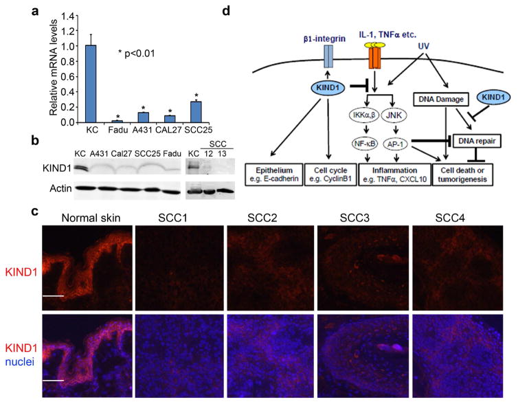
Figure 6. KIND1 is reduced in SCC cells and Tissues.
(a) qRT-PCR of KIND1 with total RNA isolated from human keratinocytes and SCC cell lines. GAPDH was used as an internal control. P-values of <0.05 were obtained via student T-test. (b) Immunoblotting for KIND1 with protein lysates isolated from human keratinocytes and SCC cell lines. Actin was used as a loading control. (c) Immunostaining of frozen tissue sections of human skin and SCC samples for KIND1 followed by detection with an Alex555 dye-conjugated secondary antibody. KIND1 [orange], nuclei [Hoechst, blue]. Scale bars: 100 μm. (d) Working model depicting multi-functions of KIND1.

