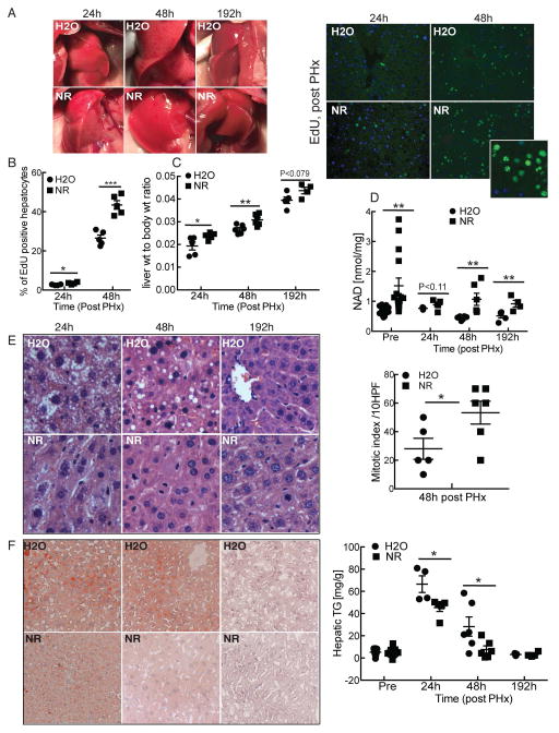
Figure 1. Nicotinamide riboside promotes liver regeneration.
10–14 week old male C57BL6/J mice were treated with nicotinamide riboside (NR) at a dose of ~500mg/kg/day. After 14 days, animals were subjected to 2/3 partial hepatectomy and analyzed 24, 48 and 192 hours later (n = 6 per group).
(A) Left: Photographs of regenerating livers. Right: EdU incorporation in regenerating livers. Inset shows an enlarged view from an NR-treated liver at 48h post PHx.
(B) Quantification of EdU positive hepatocytes.
(C) Liver to body weight ratio.
(D) Liver NAD content pre and post PHx.
(E) Left: Representative liver sections stained with H&E at 40X showing mitotic figures and micro and macro-vesicular fatty changes. Right: Quantification of mitotic figures across multiple high power fields.
(F) Left: Oil Red O staining to detect neutral lipids. Right: Hepatic triglyceride content in pre and post hepatectomized NR and H2O treated mice.
Error bars represent S.E.M. *, p < 0.05; **, p < 0.01; ***, p < 0.001.

