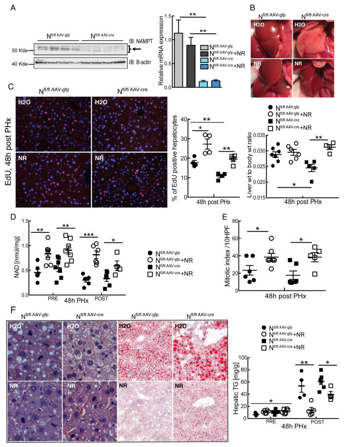
Figure 4. Hepatocyte-specific loss of Nampt impairs regeneration, and is rescued by nicotinamide riboside (NR).
Hepatocyte specific Nampt deficient mice were generated by injecting animals bearing two floxed alleles with an AAV expressing Cre recombinase (AAV-Cre). Littermates infected with AAV-Gfp served as controls. Treatment with NR (~500 mg/kg/day) began 5 days after infection and 2 weeks prior to PHx. Analyses were performed 48 hours after PHx.
(A) Protein and mRNA expression of Nampt in livers.
(B) Photographs of regenerating livers.
(C) Left: Proliferating hepatocytes identified by EdU detected by immunofluorescence (red) and counterstained with DAPI (blue). Middle: Quantification of EdU positive hepatocytes (n=4–5/group). Left: Liver to body weight ratios.
(D) NAD content in livers before and after PHx.
(E) Mitotic index as determined by counting mitotic figures in hepatocytes under high power in H&E stained sections (n= 5–6/group).
(F) Left: Representative liver sections stained with H&E (left panels) and Oil Red O (right panels). Right: Hepatic triglyceride content.
Error bars represent S.E.M. *, p < 0.05; **, p < 0.01; ***, p < 0.001.

