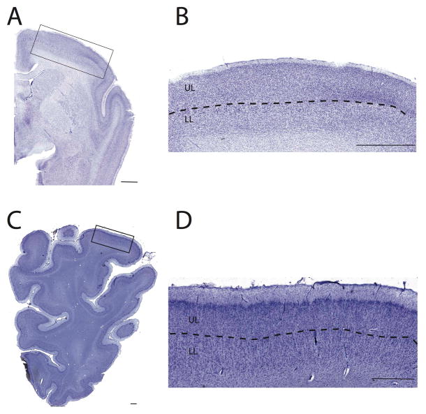
Figure 3.
Examples of delineations used to identify upper (UL) and lower layers (LL) in a pygmy marmoset (A, B), a hippopotamus (B, C). There is extensive variation in layer IV such that the presence of layer V neurons was used to distinguish upper versus lower layer neurons. In the motor cortex, upper and lower layers were distinguished by large cells located in layer V. Within other regions, such as the primary somatosensory cortex, upper layers were distinguished by the presence of granular cells located in layer IV. In some species such as the hippopotamus, layer IV was difficult to identify. The presence of large cells in layer V was used to distinguish upper from lower layers. Scale bar: 1.5 mm.

