
Figure 1.
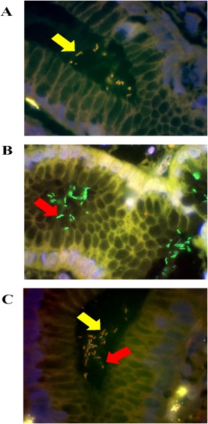
Detection of H. pylori and determination of clarithromycin susceptibility in gastric biopsies from three different patients by FISH. Probes were visualised using a triple filter. Green fluorescence indicates clarithromycin sensitive H. pylori (B and C); yellow fluorescence indicates clarithromycin resistant H. pylori (A and C). The DAPI counterstain produces blue fluorescence (A, B and C). Mixed infection is present within the same biopsy specimen in panel C. Arrows indicate the presence of H. pylori infecting the gastric mucosa (Magnification, X100).