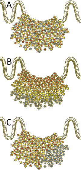
Figure 3.

Scheme of three different kinds of visualised heterogeneity in tumour cell composition of a primary tumour. (a) Mosaic‐like heterogeneity form with diffuse intermingling cells at different intensity levels. (b) During ngTMA construction, we decided to search for possible differences between the tumour centre and the invasive front, which is a classical approach to the study, eg hypoxia related effects or simply the diagnostic background of biopsies versus resection specimens. Here, highlighted as decreased intensity levels to the periphery. In this way, an assumed and experimentally targeted heterogeneity was tested. (c) Haphazard heterogeneity takes place, if certain areas (low right) are outside the usual expected spectrum of the rest of the tumour staining. This kind of heterogeneity was originally the main argument against the use of TMA for this purpose 3. However, it is currently accepted that an increased number of cores per tumour can balance this effect 23. Additionally, we show how to quantify and outline such differences and cases. Of note, any kind of haphazard heterogeneity can be transferred to a targeted heterogeneity, if, eg immunohistochemical analysis is used as the basis for ngTMA construction.
