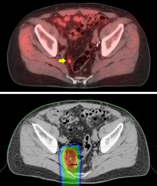
Figure 5.

72-year-old with Gleason 9 (5 + 4) and PSA 10.8. He underwent a prostatectomy and his PSA remained < 0.1 ng/mL for 7 years. PSA then began to rise, 0.63 ng/mL with a PSA doubling time of 9.3 months. An abdominal and pelvic CT as well as technetium bone scans where negative. 11C-Acetate PET/CT imaging showed a metabolic 9 mm right peri-rectal lymph node (top image, yellow arrow). A small 5 mm metabolic node was also seen higher up in the left pelvis (not shown). No metabolic lesions were seen in the prostate bed and no lesions were seen on the study to suggest distant metastatic disease. Radiation treatment to the prostate bed with radiation extending to the pelvic lymph nodes is technically viable. This case was complicated, however, by a history of ulcerative colitis, making standard radiation problematic. The patient opted to undergo Intensity Modulated Proton Therapy. The proton therapy was administered to the pelvic lymph nodes detected on the 11C-Acetate imaging alone. The 11C-Acetate images were electronically integrated into the treatment plan to help guide the proton therapy. The bottom image shows the targeting of the proton beam treatment (color areas), which is narrow and mostly avoids the colon. Two years following treatment, the PSA has remained < 0.1 ng/mL. He experienced no side effects from the radiation treatment and importantly, has not required hormonal therapy or experience exacerbation of his ulcerative colitis.
