
Figure 2.
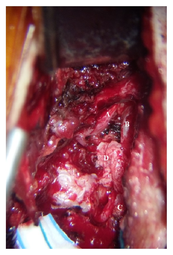
Intraoperative picture showing (A) L2 nerve root, (B) dural sac, (C) L3 nerve root, and (D) the posterior extruded segment.
Official websites use .gov
A
.gov website belongs to an official
government organization in the United States.
Secure .gov websites use HTTPS
A lock (
) or https:// means you've safely
connected to the .gov website. Share sensitive
information only on official, secure websites.
Intraoperative picture showing (A) L2 nerve root, (B) dural sac, (C) L3 nerve root, and (D) the posterior extruded segment.