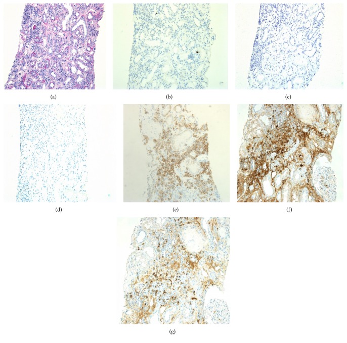
Figure 2.
Light microscopic findings for a 3-month protocol biopsy specimen. (a) Mononuclear inflammatory cell infiltration was seen in the interstitial lesions, with plasma cells accounting for approximately 20%. Moderately developed tubulitis was noted in the middle of the field (PAS, ×200). (b–d) Negative staining of SV40 (b), EBER (c), and IgG4 (d) in allograft specimen (×200). (e) CD138-positive interstitial inflammatory cells (×200). (f, g) Infiltrating plasma cells positive for both kappa (f) and lambda (g) light chains, indicating that they are polyclonal (×200).

