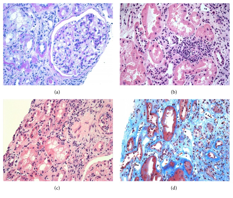
Figure 3.
Light microscopic findings in a second biopsy at 3 months after the first antirejection therapy. (a) Transplant glomerulitis, mild (PAS, ×100), (b, c) mild infiltration of inflammatory cells including plasma cells; and mildly developed tubulitis was noted (HE, ×400 (b), ×200 (c)). (d) Peritubular capillaritis was moderately developed (Masson's Trichrome, ×400).

