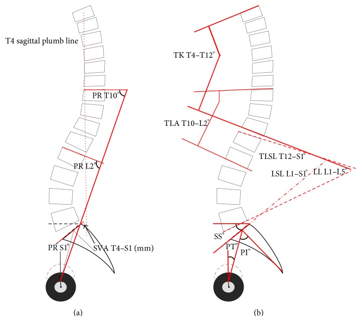
Figure 4.
Illustration of the assessed radiographic spinal outcome parameters. RKA: regional kyphosis angle; TLA (T10–L2): thoracolumbar junction angle T10–L2; PI: pelvic incidence; PT: pelvic tilt; SS: sacral slope; TLSL T12–S1: thoracolumbosacral lordosis T12–S1; LSL L1–S1: lumbosacral lordosis L1–S1; LL L1–L5: lumbar lordosis L1–L5; TK T4–T12: thoracic kyphosis T4–T12; PR-S1: pelvic radius to S1; PR-T10: total lumbopelvic lordosis to T10; PR-L2: regional lumbopelvic lordosis to L2; SVA T4–S1: sagittal vertical axis T4 to S1.

