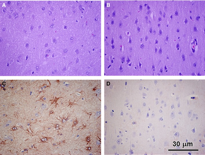
Figure 1.

Light micrographs of paraffin sections of the cerebral cortex external granular layer from the neuronal ceroid lipofuscinosis‐affected Cane Corso (A and C) and from an age‐matched normal Beagle (B and D). A and B were stained with hematoxylin and eosin. C and D were immunostained with an anti‐GFAP antibody. The diseased dog exhibited a substantial reduction in cell density in the external granular layer and substantial numbers of GFAP‐labeled astrocytes that were not observed in the control dog sample. Bar in (D) indicates magnification of all micrographs.
