Figure 2.

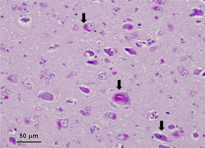
Light micrographs of diastase‐treated PAS‐stained section of the cerebral cortex from the affected dog. Aggregates of PAS‐staining inclusions were present in the perinuclear areas of most cortical neurons (arrows).

Light micrographs of diastase‐treated PAS‐stained section of the cerebral cortex from the affected dog. Aggregates of PAS‐staining inclusions were present in the perinuclear areas of most cortical neurons (arrows).