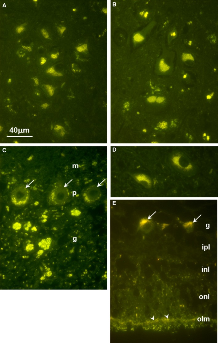
Figure 4.

Fluorescence micrographs of unstained sections of (A) cerebellar cortex, (B) cerebral cortex, (C) retina, (D) deep cerebellar nucleus, and (E) retina. In (A), arrows point to Purkinje cells; m: molecular layer, p: Purkinje cell layer, g: granular cell layer. In (C), arrows point to Purkinje cell bodies. In (E), arrows point to retinal ganglion cells and arrow heads point to the outer limiting membrane; g: ganglion cell layer, ipl: inner plexiform layer, inl: inner nuclear layer, onl: outer nuclear layer. Bar in (A) indicates magnification of all micrographs.
