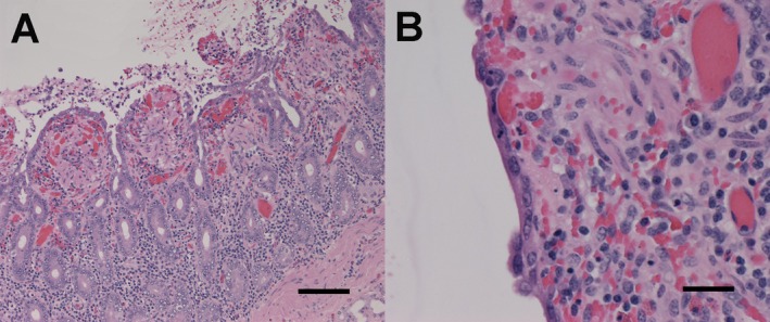
Figure 2.

Microscopic lesions in the duodenum of a horse inoculated with Clostridium difficile toxins. In (A), there is blunting of villi with loss of epithelium lining villous tips. The lamina propria is expanded by congestion and hemorrhage. The lumen contains inflammatory cells intermixed with sloughed epithelial cells, erythrocytes, and fibrin. Magnification 10×, hematoxylin and eosin (H&E) stain. In (B), the intact epithelial cells are flattened and stretched across the surface of an affected villus. Magnification 40×, H&E stain. Lesions presented are from horse 5. Figure A length of the scale bar = 100 μm. Figure B length of the scale bar = 25 μm.
