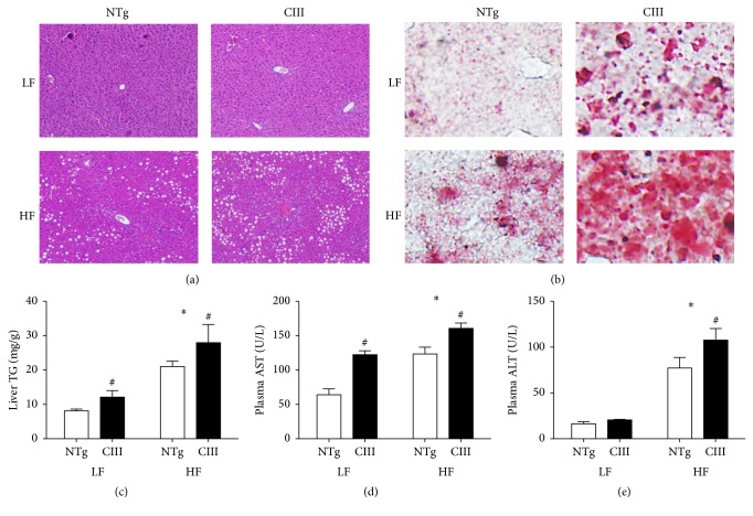
Figure 1.
Overexpression of apoCIII promotes hepatic steatosis and dysfunction. NTg and apoCIII mice fed with low-fat (LF) or high-fat (HF) diets for 16 weeks. Representative liver sections isolated from mice stained with (a) HE and (b) Oil Red O. Note the macrovesicular lipid deposits, which appear as white spots in the HE-stained tissues (a), and the presence of lipids, which are stained in red (b). (c) Liver triglyceride content determined by enzymatic assay (n = 7-8). (d) Plasma concentrations of the transaminases ALT and AST (n = 5). Data are expressed as the mean ± SEM. ∗LF versus HF groups; #NTg versus apoCIII mice (p < 0.05; two-way ANOVA).

