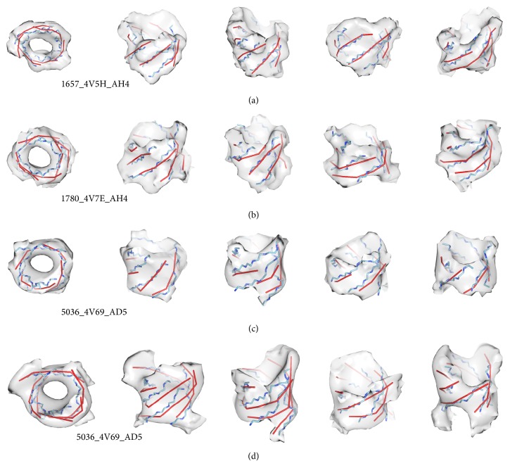
Figure 7.
β-Traces detected from β-barrel density maps obtained from experimentally derived cryo-EM density maps. The best of the fifteen sets of modeled β-traces (red) are superimposed with the backbone of the β-strands (blue) and the density maps (gray) for β-barrel 1657_4V5H_AH4 (EMD_1657, sheet AH4 of protein 4V5H) in (a) and 1780_4V7E_AH4 (EMD_1780, sheet AH4 of protein 4V7E) in (b). The top view (left) and four side views are shown in each case. (c) A conservative segmentation for β-barrel 5036_4V69_AD5 (EMD_5036, sheet AD5 of protein 4V69) and its corresponding β-strands modeling result. (d) A relaxed segmentation of the same β-barrel as in (c) superimposed with the backbone and the detected β-traces. β-Barrels in (c) and (d) are shown using the same density threshold in similar view points.

