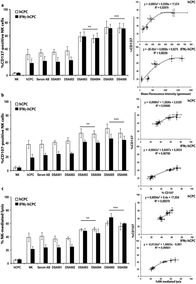
Figure 5. DSA-HLA I-A2 of high and intermediate binding strength induce ADCC in hCPC.
IL-15-activated NK cells were cultured alone or with HLA-A2 positive hCPC or IFNγ-hCPC (n = 4) in the presence of control serum AB or DSA-HLA-I-A2 sera (DSA001-006). (a) % CD137-positive NK cells, (b) % CD107-positive NK cells, and (C) % NK cell-mediated lysis evaluated as % 7AAD-positive hCPC. Results represent mean values ± SD from four different experiments of each hCPC. The percentages of CD137-positive NK cells with hCPC or IFNγ-hCPC were plotted as function of respective MFIs of DSA-HLA-I-A2 sera (upper right panel) or as function of % CD107-positive NK cells (middle right panel), the % CD107-positive NK cells were plotted as function of % NK-mediated lysis (low right panel) for both hCPC and IFNγ-hCPC. Statistical analyses were performed using Mann–Whitney test for non-paired groups. **P < 0.01 and ***P < 0.001 compared to NK + hCPC.

