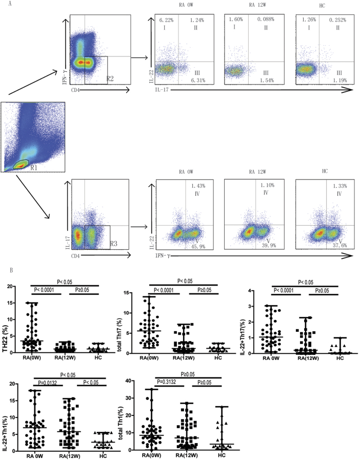
Figure 2. The percentages of circulating Th22, total Th17, and IL-22+Th17, IL22+Th1, but not total Th1 cells decreased following MTX+LEF therapy only in patients that exhibited clinical improvement (n = 40).
Peripheral blood mononuclear cells (PBMCs) were collected from RA patients responsive to treatment at baseline (before treatment, 0 week) and after treatment (12 weeks) with MTX+LEF, and analyzed by flow cytometry for the percentage of different Th subsets, including IFN-γ−IL-17−IL-22+ (Th22), IFN-γ−IL-17+ (total Th17), IFN-γ−IL-17+IL-22+ (IL-22+Th17), IFN-γ+IL-17− (total Th1), and IFN-γ+IL-17−IL-22+ (IL-22+Th1) cells. (A) The gating strategy for detecting different subsets of Th cells and representative flow images on samples from RA patients at 0 week (0 W), 12 week (12 W), or from healthy controls (HC; n = 20). R1, lymphoctyes; R2, IFN-γ−CD4+T cells; R3, IL-17−CD4+T cells; I, Th22 cells; II, IL-22+Th17 cells; III, IL-22−Th17 cells; IV; IL-22+Th1 cells; V; IL-22−Th1 cells.

