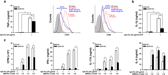
Figure 2. NLRP3 inflammasome activation in IgA ICs-primed dendritic cells.
TNF-α secretion measured by ELISA, expression levels of CD40+ and CD86+ (within gated CD11c+ cells; the gray-filled area created by staining with an isotype-matched control antibody) determined by flow cytometry in BMDCs from untreated wild type or NLRP3 KO mice, which were incubated for 24 h with IgA ICs (a). IL-1β secretion measured by ELISA in BMDCs from untreated wild type or NLRP3 KO mice, which were incubated for 24 h with IgA ICs and 30 min ATP (b). T cell proliferation was measured by [3 H]-thymidine, and secretion of IFN-γ, IL-17A and IL-4 by ELISA at the indicated ratio of BMDCs:T cells for 3 days, in which BMDCs were incubated for 24 h with IgA ICs and cocultured with OT-II CD4+ T cell pulsed with OVA peptide (c). The data are expressed as the mean ± SEM for three separate experiments. *p < 0.05. WT, wild type. #Not detectable. BMDCs, bone marrow derived dendritic cells. IgA ICs, IgA immune complexes.

