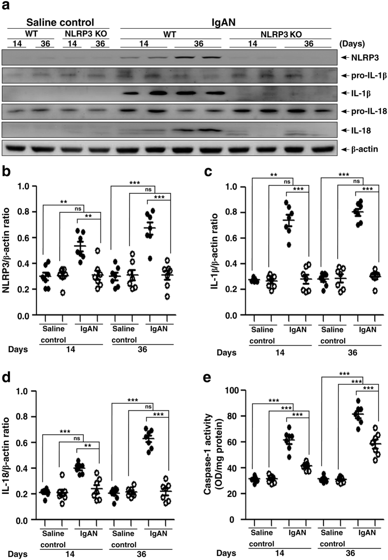
Figure 5. Renal NLRP3 inflammasome activation in wild type or NLRP3 KO mice with IgAN.
Representative Western blots of NLRP3, IL-1β, or IL-18 levels in kidney tissues on day 14 and day 36, β-actin was used as the loading control (a); semi-quantification of the NLRP3/β-actin ratio (b), IL-1β/β-actin ratio (c), or IL-18/β-actin ratio (d). Renal caspase 1 activity (e). The data are expressed as the mean ± SEM for 7 mice per group. **p < 0.01; ***p < 0.005. WT, wild type. ns, no significant difference.

