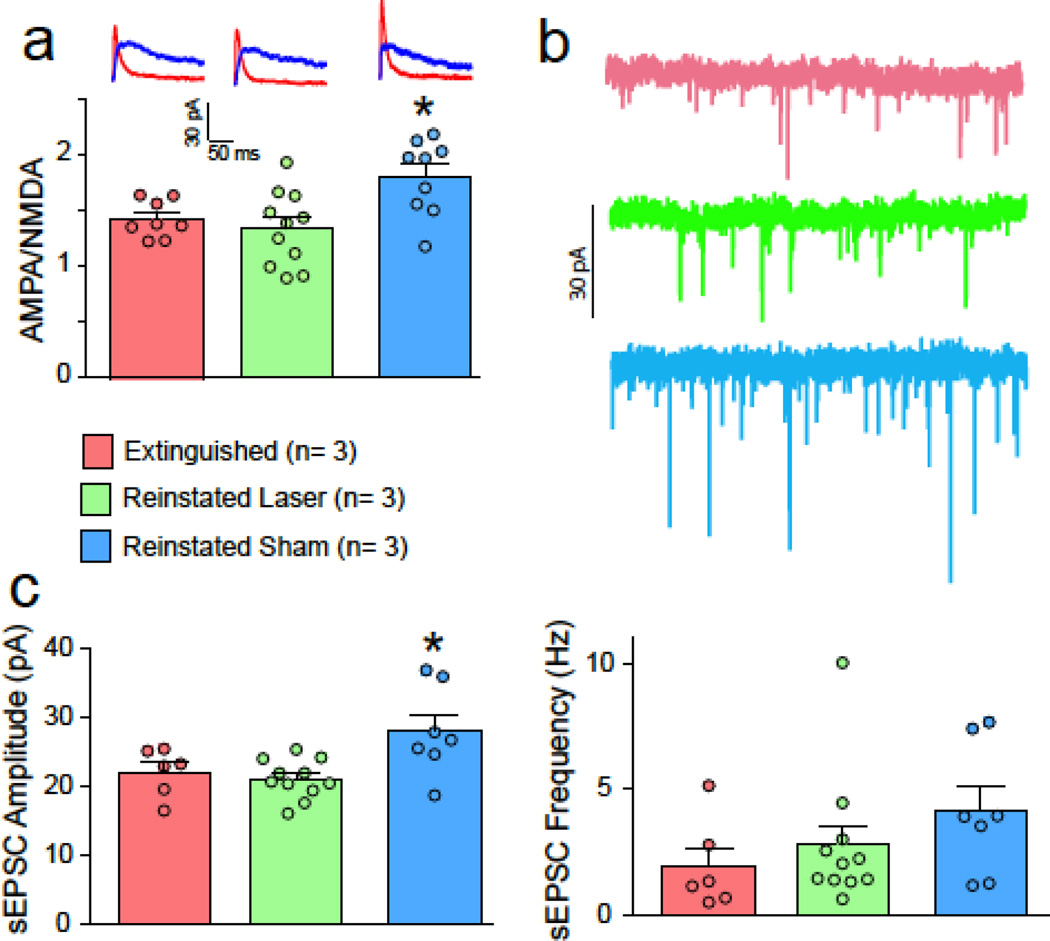
Figure 3. Optical inhibition of PL-to-NAcore projection prevents synaptic potentiation.
A) Increase in AMPA/NMDA after cue-induced reinstatement was blocked by 15 min optogenetic inhibition of PL terminals in the NAcore. Data shown as mean ± SEM ratio for each treatment group, with individual cells indicated in smaller circles. N refers to number of animals. Representative traces are shown for each treatment in the left panel. B) sEPSC amplitude was increased in the sham but not the laser group, supporting a postsynaptic potentiation mechanism. C) sEPSC frequency was not different between groups.
*p< 0.05, comparing reinstated sham to the other groups.

