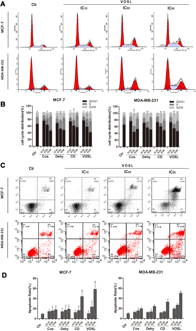
Figure 6. Effects on cell cycle and apoptosis of breast cancer cells after Cos, Dehy, CD, or VOSL treatment.
MCF-7 cells or MDA-MB-231 cells were planted into 6-well plates at 3 × 10⁵ cells/well, incubated with the respective IC10, IC30, IC50 concentrations of Cos, Dehy, CD, or VOSL for 48 h, cells were harvested by trypsinisation, and then fixed by ice-cold ethanol (70%). After washing with PBS, the cell pellets were resuspended in propidium iodid (PI) staining buffer (50 μL/mL PI, RNase A). After 15 min of incubation at 37 °C, cell cycle distribution was analyzed by a FACScalibur System using ModFit software (A,B). MCF-7 or MDA-MB-231 cells were planted into 6-well plates at 2 × 10⁵ cells per well, treated with the respective IC10, IC30, IC50 concentrations of Cos, Dehy, CD, or VOSL for 48 h, stained with Annexin V-FITC/PI, and then detected by a FACScalibur system (C,D). The cell cycle distribution and apoptotic percentages from three independent experiments were analyzed and compared, *p < 0.05 and **p < 0.01 compared with the control group.

