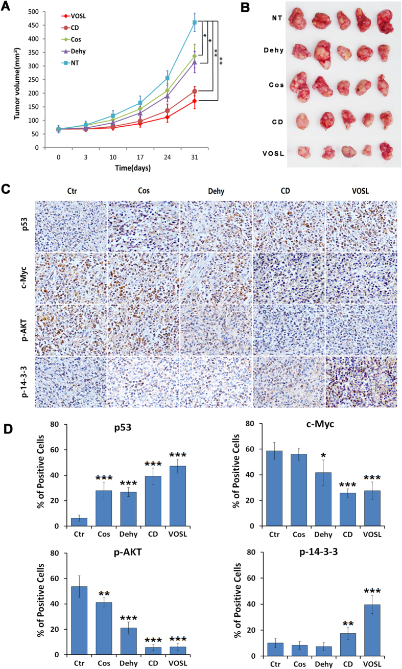
Figure 7. VOSL and its main active ingredients suppress the growth of breast cancer MDA-MB-231 xenografts.
(A,B) The xenograft mouse models were randomly divided into five groups. The Cos, Dehy, CD and VOSL-treated groups were injected intraperitoneally at a dose of 20 mg/kg/day, respectively. The negative control (NT) was treated with an equal volume of vehicle. Tumor size was monitored at 0, 3, 10, 17, 24 and 31 days post-treatment and compared at 31 days post-treatment; *p < 0.05 and **p < 0.01 compared with the negative control (NT) group. (C,D) Tumor-bearing mice were sacrificed after 30 times of administrations and tumors were harvested and weighed, and then were cut into consecutive sections for examining the expression of p-AKT, p53, p-14-3-3 and c-Myc by immunohistochemistry. Original magnification 200×. The positive cells of the relevant factors in xenografts were presented as mean ± SD, *p < 0.05 and **p < 0.01 compared with the NT control group.

