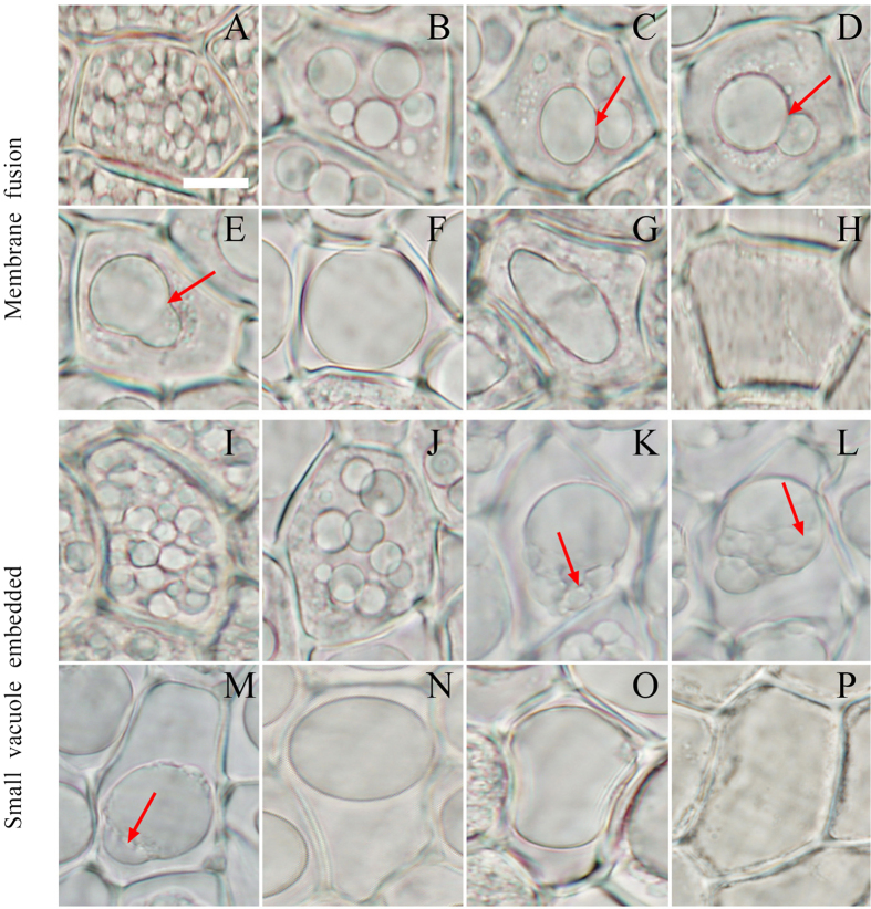
Figure 1. The integration and transformation of vacuoles in the PCD of aleurone layers.
The aleurone layers stripped from grains cultured for different times were observed using fluorescent microscopy, and the vacuoles exhibited two types of fusion. (A–H) Vacuole fusion through membrane fusion; arrows indicate the mutual contact section (C,D) and the mutual fusion section (E) of the tonoplast. (I–P) Vacuole fusion through embedded fusion; arrows indicate the small vacuole starting to enter the larger vacuole (K), the small vacuole in the larger vacuole (L), and the trace of the small vacuole in the larger vacuole (M). The assay was performed at least three times and a representative aleurone layer section is shown. The bar represents 10 μm.

