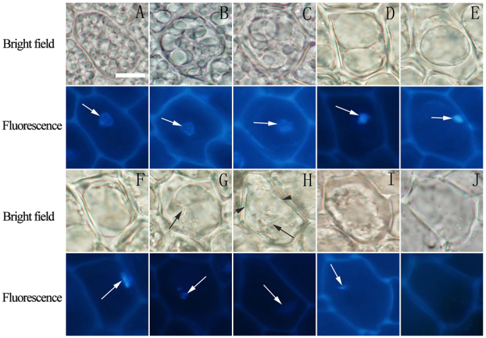
Figure 5. Nuclear morphological changes of aleurone cells at different stages of cell death.

The morphology of nuclei in the aleurone cells stained with 1 μg mL−1 DAPI was observed by fluorescence microscopy. (A–J) The nuclei stained with blue fluorescence DAPI (arrows) and the corresponding images under bright field. Both experiments were repeated, and similar images were obtained. The bar represents 10 μm.
