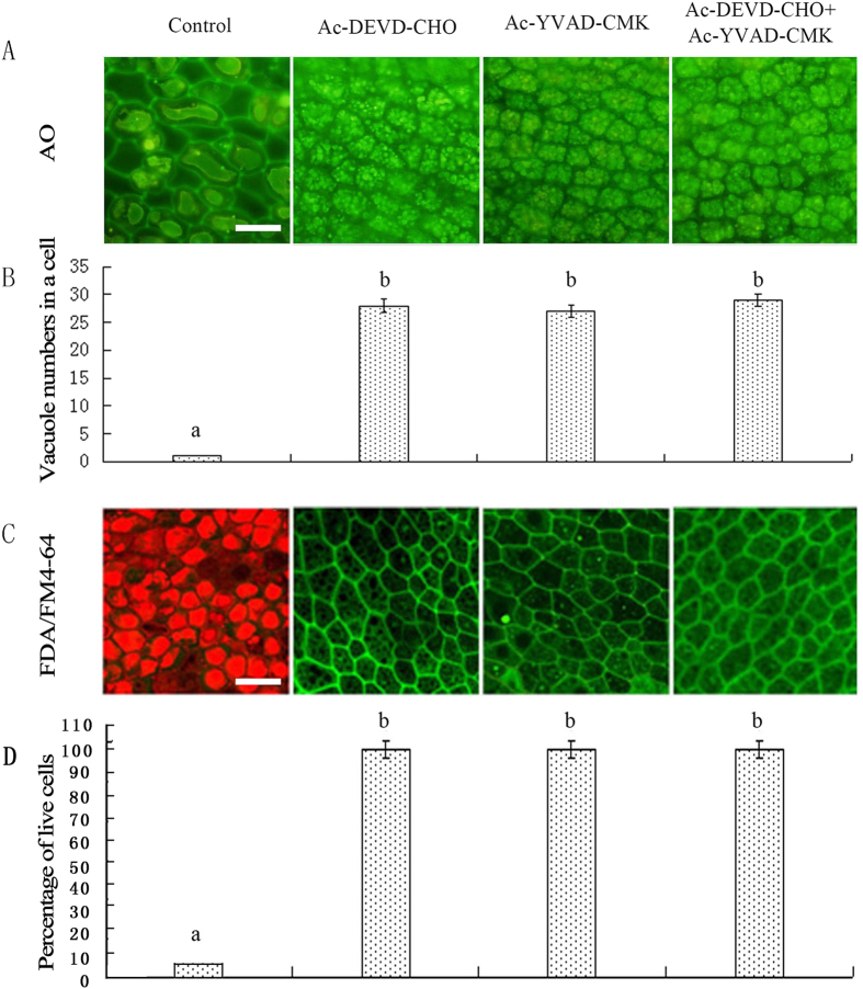
Figure 7. Ac-YVAD-CMK and Ac-DEVD-CHO slow the process of vacuolar coalescence.
Rice aleurone layers were isolated from seeds imbibed for 2 d and then incubated with distilled water, 100 μM Ac-DEVD-CHO, 100 μM Ac-YVAD-CMK or 100 μM Ac-DEVD-CHO plus 100 μM Ac-YVAD-CMK for 7 d. Finally, the layers were examined for vacuolation after 8.5 μg mL−1 AO or 2 μg L−1 FDA/1 μg L−1 FM4-64 staining using LSCM. (A) Live cells exhibit AO green fluorescence. (B) Statistical analyses were conducted on the vacuole number per cell. (C) The live cells exhibit FDA green fluorescence and the dead cells exhibit FM4-64 red fluorescence. (D) Viability of the cells was quantified from at least four aleurone layers. The assay was performed at least three times, and a representative aleurone layer section is shown. Data represent the mean ± SD from three independent biological replicates, and different letters indicate a significant different at p < 0.05 according to Duncan’s multiple range test. The bar represents 50 μm.

