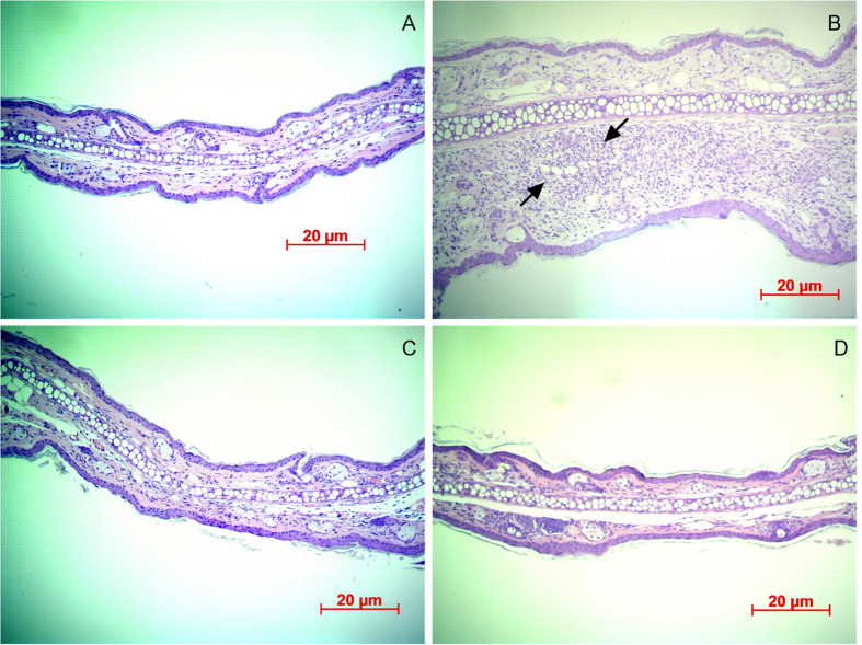
Figure 7. Histological observations of ears of ICR mice with hematoxylin and eosin staining.
(A) Original ear. (B) Increases were observed in both the ear thickness and the number of infiltrating inflammatory cells (arrows) surrounding the injection site of P. acnes. The ear thickness, swelling, erythema, and inflammatory reactions were reduced (C) in LY-shelled-MBs-treated ears and (D) in ears treated with both US and LY-shelled MBs.

